Imaging Hardware and Applications
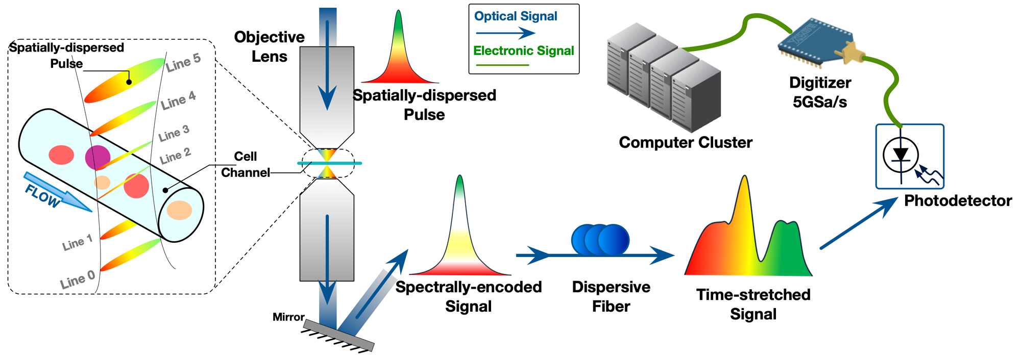
Recent advances in high-throughput cell microscopy promise a new generation of image-based biomedical applications, e.g., cell sorting and classification, otherwise impossible with traditional biomolecular assays. As the prior art in the cytometry technique, ATOM system (as illustrated in FIG.1) is a home-built optical microscopy for cell flow imaging by our colleborator. One challenge with this system, however, is that we must derive adequate information from the cell images in realtime to facilitate various forms of image analytics while avoiding significant impact on the overall imaging throughput.
As FIG.1, the cell flow imaging adopts the line scan technique, where the periodic laser pulse shines through the cell tunnel, and each pulse encodes an image line to the time-stretched signal. Subsequently, a digitizer (ADC) samples the continuous signal in an ultra-high-speed (5 Giga samples per second), which still limits the imaging resolution. Hence, this research investigates a realtime super-resolution (SR) scheme for the ATOM system under the speed limitation of ADC.
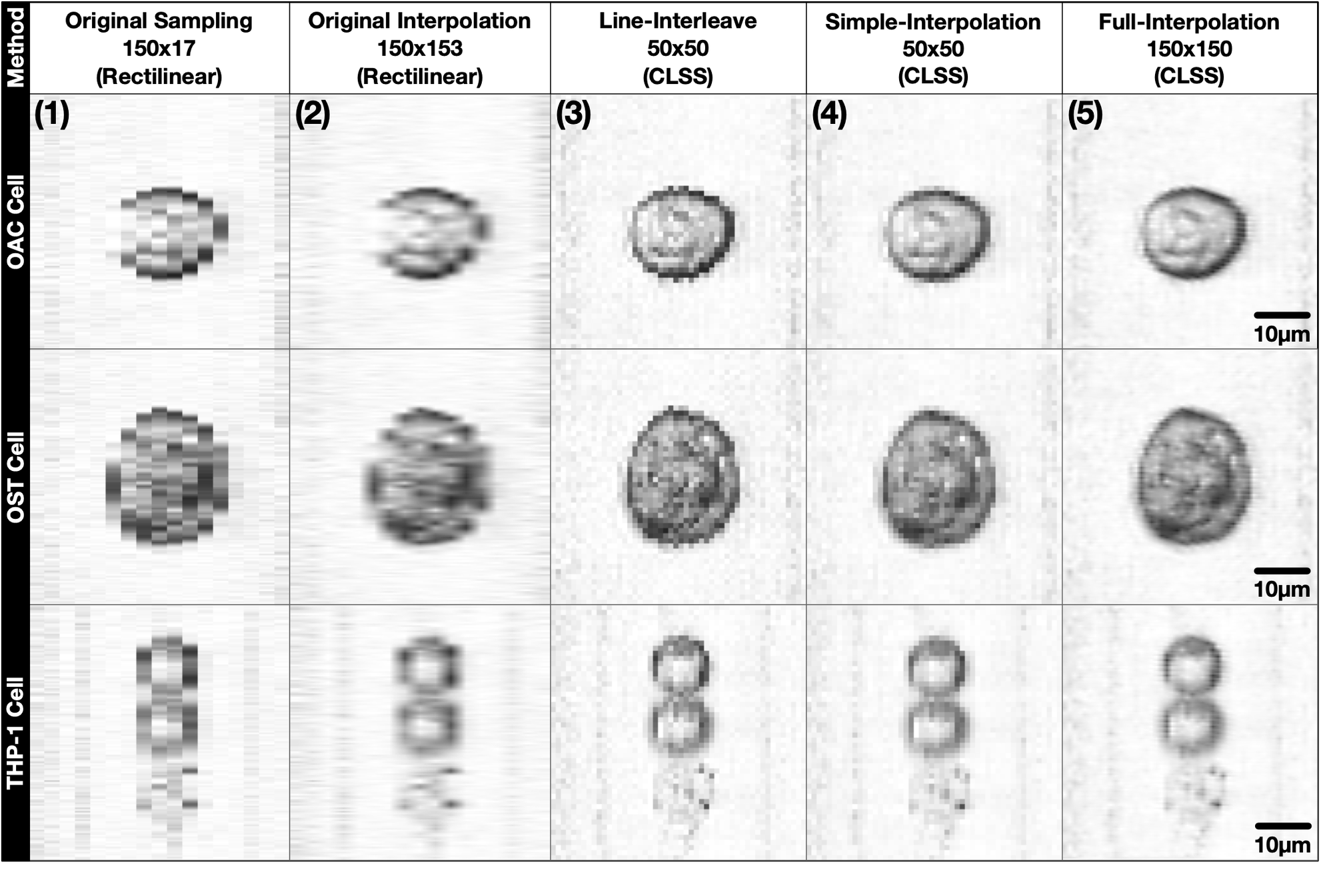
We proposed a novel technique, coprime line scan super-resolution (CLSS), for not only ATOM microscopy but also imaging systems with a similar mechanism. Further, we designed FPGA based hardware architecture for CLSS that reaches a realtime processing throughput of 5 Gigapixels per second, which is scarcely possible on other platforms. The realtime super-resolution on FPGA facilitates the subsequent image-based cell classification and sorting. FIG.2 demonstrates cell images for comparison. These images were sampled with the same ADC frequency, but different reconstruction schemes: (1) original sampling without SR; (2) with conventional rectilinear SR (interpolation); (3)-(5) with different options in CLSS sampling scheme.